


Read More


The 5cm lift: Activating your entire body's muscles
08-05-2026 12:00 HKT


Mother's Day dining revenue drops $50m, expert says
11-05-2026 13:30 HKT

Hong Kong’s iconic Lamma Winds decommissioned after two decades
11-05-2026 18:07 HKT
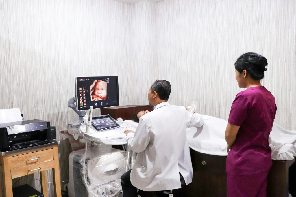
Ultrasound examination is a commonly used tool in modern medicine, widely applied from prenatal checks to internal organ assessments. However, various misunderstandings and concerns about ultrasound still circulate publicly. Some people worry about radiation causing cancer, while others mistakenly believe it is only for pregnant women. Dr. Vivian Leung Ji-fong, Assistant Professor at the School of Nursing and Health Studies, Hong Kong Metropolitan University, helps debunk 7 common ultrasound myths for everyone.
Ultrasound examination uses high-frequency sound waves to produce real-time images, allowing for safe and quick visualization of internal body structures and tissues. Because it does not involve ionizing radiation and is versatile, it is widely used in various clinical diagnoses.
Many people confuse ultrasound with X-rays or CT scans, worrying it might damage cells or cause cancer. In fact, ultrasound uses high-frequency sound waves that reflect off body tissues to form images; the process involves no ionizing radiation. According to international research, as long as it is operated according to safety guidelines, diagnostic ultrasound does not cause genetic damage or induce cancer. It remains safe even when needed for long-term monitoring of chronic diseases.
Claims circulating online that "too many ultrasounds during pregnancy can harm the baby" have no scientific basis. Medical ultrasound is a safe and reliable prenatal examination tool that poses no risk to the fetus. Conversely, it helps in the early detection of congenital abnormalities (such as heart or neural tube defects), allowing the medical team to intervene and follow up early, ensuring the safety of both mother and baby. In decades of clinical application, no evidence has shown a causal link between ultrasound and fetal abnormalities or autism.
The vast majority of ultrasound examinations are non-invasive procedures. The medical professional simply applies a small amount of gel to the skin and gently glides the probe over the area. Most people only feel mild pressure or a cool sensation and do not experience pain. As for some endoscopic examinations, such as transvaginal or transrectal ultrasounds, they might cause mild discomfort. However, medical professionals explain the procedure beforehand and strictly ensure privacy, guaranteeing safety and peace of mind.
Although ultrasound can display internal conditions in real-time, its accuracy can be affected by several factors, such as the operator's skill, the patient's body size, interference from intestinal gas, and even fetal position. Ultrasound provides information on the morphology and blood flow of lesions, but it cannot be used alone to determine whether a tumor is benign or malignant. Doctors need to combine medical history, blood tests, MRI scans, or biopsy results to make a final accurate diagnosis.
Many people think "ultrasound equals baby scans," but obstetric applications are just the tip of the iceberg. Today, ultrasound is widely used for examining the whole body, such as checking the liver, gallbladder, kidneys, gallstones, fatty liver, heart valves, vascular stenosis, and deep vein thrombosis. In sports medicine, doctors also use its dynamic imaging to observe tendon tears and assess ligament recovery.
Although ultrasound is very safe, the medical field always emphasizes the principle of "performing examinations based on clinical need." Excessive testing not only wastes medical resources but might also detect "incidentalomas" that pose no malignant risk, on the contrary causing unnecessary anxiety and follow-up for the patient. Prenatal and clinical guidelines have clear recommendations on the frequency of scans. Good health management should be based on a balanced diet and moderate exercise, not blindly increasing the number of tests.
Portable ultrasound scanners are becoming more common, but professional image interpretation is by no means simply "looking at the picture." The operator must possess in-depth knowledge of anatomy, skills in adjusting imaging parameters, and the ability to identify pathology. Non-professionals using these devices are highly prone to misdiagnosis, which can delay treatment. In Hong Kong, qualified operators must undergo professional certification, continuous education, and quality monitoring to ensure patient safety.
Ultrasound is a safe, non-invasive, real-time imaging tool that can assist doctors in early diagnosis and disease monitoring. As long as the public masters the correct medical knowledge and discusses examination needs rationally with doctors, ultrasound can maximize its clinical value, and medical resources can be more effectively utilized for those who truly need them.
Author: Dr. Vivian Leung, Assistant Professor, School of Nursing and Health Studies, Hong Kong Metropolitan University
Expert Profile: Leung is a registered radiographer with many years of clinical and professional training experience in ultrasound. Her expertise includes abdominal, obstetric, pediatric, and vascular ultrasound, as well as liver elastography. She is actively involved in teaching and research, authoring books and journal articles, and lecturing at local and international conferences, nurturing the next generation of ultrasound professionals.