


Read More
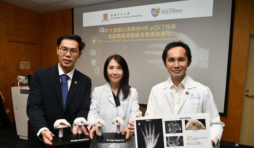
The Faculty of Medicine at the Chinese University of Hong Kong (CU Medicine) has used high-resolution scanning technology to analyze bone erosion patterns in rheumatoid arthritis, offering hope for earlier intervention and reducing the risk of permanent joint damage.
ADVERTISEMENT
SCROLL TO CONTINUE WITH CONTENT
Rheumatoid arthritis is a chronic autoimmune disease affecting around 30,000 people in Hong Kong. Bone erosion, which often occurs in the metacarpophalangeal joints, is an important marker for assessing disease severity and predicting irreversible joint damage.
Conventional imaging scans have limited sensitivity for detecting early or minute erosion, hindering prompt diagnosis during the crucial first two years after onset. To address the issue, the CU Medicine research team conducted an eight-year study using high-resolution peripheral quantitative computed tomography (HR-pQCT).
They analyzed bone scans of the second metacarpal head from 247 rheumatoid arthritis patients and compared them with scans from 78 healthy individuals. This approach showed high precision in distinguishing non-inflammatory, minor bone defects from true, subtle erosive lesions.
The results indicate that HR-pQCT can help physicians accurately diagnose the condition within the “golden window” of treatment. Early initiation of treat-to-target therapy in newly diagnosed patients can also help reverse large-scale bone erosions.
Tam Lai-shan, professor at the Department of Medicine and Therapeutics and a rheumatology specialist, urged people to seek medical attention promptly if they experience persistent joint swelling, morning stiffness, or hand pain.
She said the research team plans to integrate HR-pQCT into early assessment protocols and develop an imaging-based risk stratification model to identify high-risk individuals before clinical symptoms appear.